Perthes Disease
PERTHES DISEASE
Perthes disease is a rare childhood condition that affects the hip. It occurs when the blood supply to the
rounded head of the femur (thighbone) is temporarily disrupted. Without an adequate blood supply, the bone
cells die, a process called avascular necrosis.
Although the term “disease” is still used, Perthes is really a complex process of stages that can last
several years. As the condition progresses, the weakened bone of the head of the femur (the “ball” of the
“ball-and-socket” joint of the hip) gradually begins to break apart. Over time, the blood supply to the
head of the femur returns and the bone begins to grow back.
Treatment for Perthes focuses on helping the bone grow back into a more rounded shape that still fits into
the socket of the hip joint. This will help the hip joint move normally and prevent hip problems in
adulthood.
The long-term prognosis for children with Perthes is good in most cases. After 18 mo
nths to 2
years of
treatment, most children return to daily activities without major limitations.
SYMPTOMS:
One of the earliest signs of Perthes is a change in the way your child walks and runs. This is often most
apparent during sports activities. Your child may limp, have limited motion, or develop a peculiar running
style. Other common symptoms include:
Pain in the hip or groin, or in other parts of the leg, such as the thigh or knee (called “referred
pain.”).
Pain that worsens with activity and is relieved with rest.
Painful muscle spasms that may be caused by irritation around the hip.
Depending upon your child’s activity level, symptoms may come and go over a period of weeks or even months
before a doctor visit is considered.
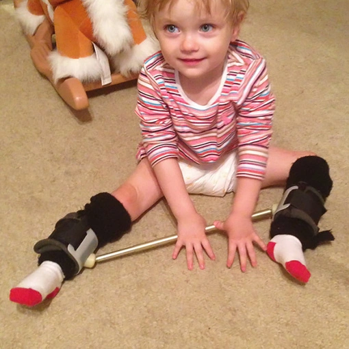
SURGICAL TREATMENT :
Keeping the femoral head within the rounded acetabulum may help the bone grow into a functional shape.
Nonsurgical treatment has not kept the hip in the correct position for healing.
The most common surgical procedure for treating Perthes disease is an osteotomy. In this type of
procedure, the bone is cut and repositioned to keep the femoral head snug within the acetabulum. This
alignment is kept in place with screws and plates, which will be removed after the healed stage of the
disease.